
BCIP-NBT Solution (Ready To Use)
A ready to use solution of chromogenic substrate of alkaline phosphatase (AP). It can be used in a wide range of applications, such as detection of alkaline phosphatase (AP) bound by antigen-antibody reaction and endogenous
alkaline phosphatase (AP) of stem cells and osteoblasts, in blotting and tissue or cell staining.
- Ready to use
- Can be used in wide range of applications such as immunochemical staining or endogenous AP staining.
- Can be used in in situ hybridization (ISH), as it is tested for DNase and RNase.
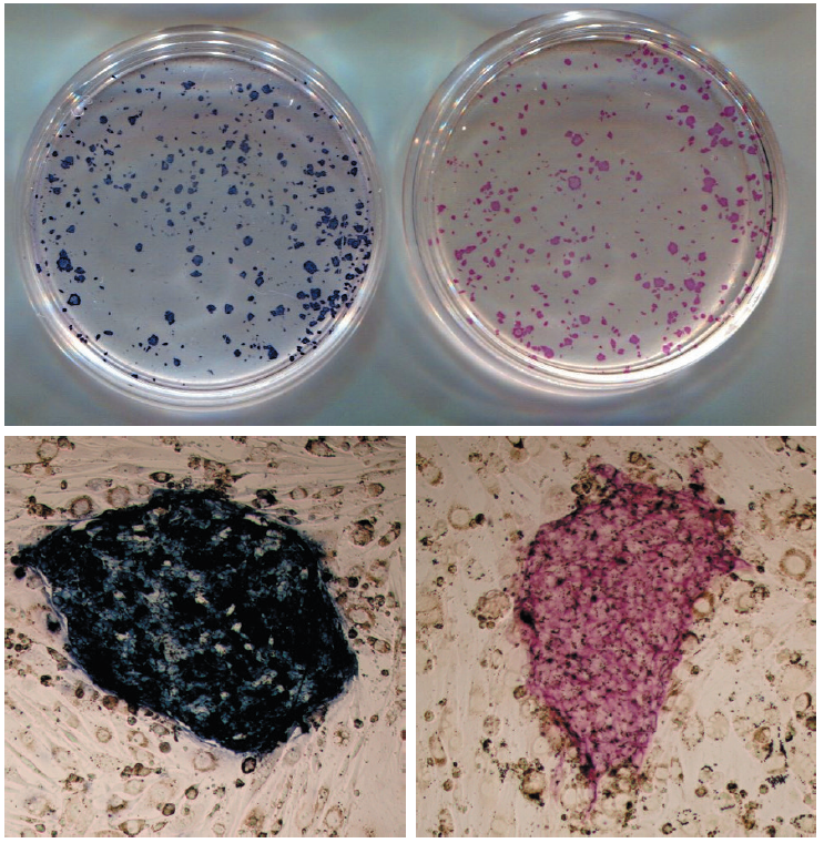
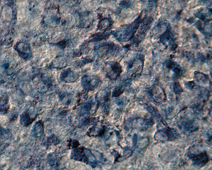
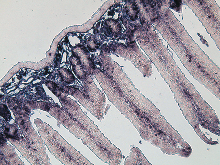
Application 1: Endogenous AP staining of iPS cells and osteoblasts
iPS cell
| BCIP-NBT Solution | Company A Red AP chromogenic substrate |
|---|---|
 |
|
| Experiment conditions |
|
|---|---|
| Cell : | iPS cell (01B7 line*), feeder cell (SNL) |
| Fixation : | 4%-Paraformaldehyde Phosphate Buffer Solution (#09154) 10 minutes at room temperature |
| Staining : | 60 minutes at room temperature |
| Detection : | (Upper) Scanner (Seiko Epson) (Lower) Optical microscope (Olympus #BX50), 10x magnification |
| *Takahashi, K. et al. Cell. 2007, 131(5), p. 861-872. | |
Osteoblasts
| Added BMP-2 (100 ng/mL) Cultured for 3 weeks |
No BMP-2 Cultured for 3 weeks |
|---|---|
 |
 |
| Experiment conditions |
|
|---|---|
| Cell : | MC3T3-E1 Cell |
| Fixation : | 4%-Paraformaldehyde Phosphate Buffer Solution (#09154) 10 minutes in room temperature |
| Staining : | BCIP-NBT Solution (Ready To Use), 30 minutes at room temperature |
| Detection : | Optical microscope (Olympus # BX50) by differential interference, 50x magnification |
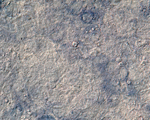
Application 2: Western blotting detection of UCP1 protein from mouse brown adipose tissue
| Company D BCIP-NBT (Substrate Set) |
BCIP-NBT Solution |
|---|---|
 |
|
| Experiment conditions |
|
|---|---|
| Sample : | Protein from mouse brown adipose tissue (From left 30 μg, 10 μg, 3 μg, 1 μg) |
| Blotting : | 5% skim milk/tTBS at room temperature for 1 hour. |
| Primary antibody : | Anti-UCP1 antibody (Rabbit) (Self-preparation) 2,000x diluted Diluted using 5% skim milk/tTBS, refrigerated overnight. |
| Secondary antibody : | Anti-Rabbit IgG (Fc), AP Conjugate(Promega #S373B) 5,000x diluted Diluted using 5% skim milk/tTBS, left at room temperature for 1 hour. |
| Detection : | 1 minute for each staining solution. |
【User evaluation】
BCIP-NBT (Substrate Set) from company D is 2 bottles type which takes time to making our own buffer. On the order hand,
1 bottle type of BCIP-NBT Solution (Ready To Use) is convenience and results as good as others.
Data reference: Professor Goto, Tsuyoshi, Department of Food and Biological Sciences, Graduate School of Agriculture, Kyoto University.
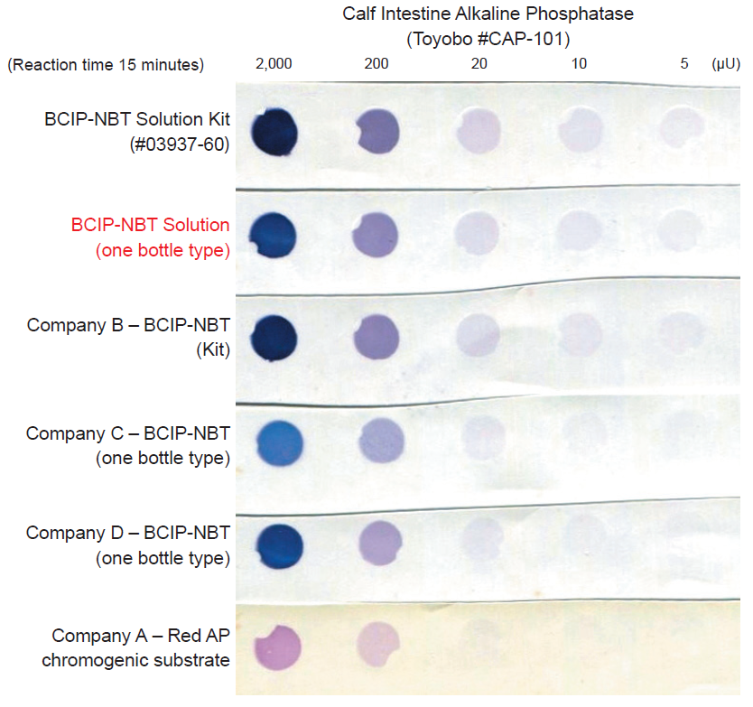
Comparison: Detection sensitivity by dot blotting
Compare with other brands, BCIP-NBT Solution has equal or better sensitivity.
Application 3: In situ hybridization (ISH)
Detecting IX type collagen (α1) mRNA from fetal mouse paraffin embedded section. The product can be used for high sensitivity detection, such as is required with in situ hybridization (ISH).
| Antisense strand | Sense strand |
|---|---|
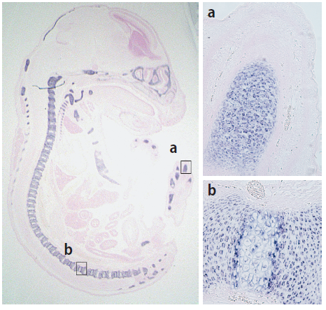 |
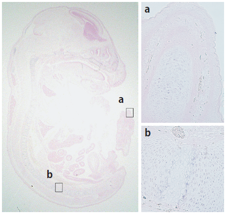 |
| Experiment conditions |
|
|---|---|
| Tissue : | Paraffin section from fetal mouse (16.5 days) for ISH (Genostaff #SMPS-08-5) |
| Hybridization : | RNA probe for DIG tag mouse Col9a1 ISH (Genostaff #MP-C-023) |
| Blocking : | Blocking One Histo (#06349-64) at room temperature for 10 minutes |
| Primary antibody : | Anti-Digoxigenin-AP, Fab fragments from sheep (Roche #11093274910) 1:2,000 Diluent: tTBS / Blocking One Histo in 19:1 ratio. |
| Staining : | BCIP-NBT Solution (Ready To Use), 4 hours at room temperature; Counterstain: Kernechtrot dye, 10 seconds at room temperature. |
| Observation : | Enclosed by CC/Mount (Diagnostic BioSystems #K002) and Entellan® new (Merck #107961), observed by Zoom Stereomicroscope MF2.6x (AXEL #1-1925-02), microscope MF 50x (Olympus #BX50) |
Application 4: Immunohistochemical staining
Detecting PCNA from mouse small intestine paraffin embedded section. If there is insufficient sensitivity, extending reaction time can result in good sensitivity.
Reaction time
5 minutes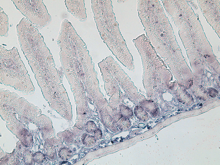 |
10 minutes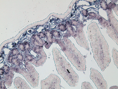 |
15 minutes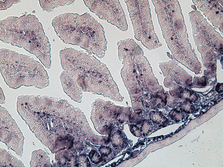 |
30 minutes
 |
| Experiment conditions |
|
|---|---|
| Tissue : | Small intestine paraffin section from mouse |
| Antigen activation : | HistoVT One (10x, pH 7.0) (#06380) at 90°C for 20 minutes |
| Blocking : | Blocking One Histo (#06349-64) at room temperature for 10 minutes |
| Primary antibody : | PCNA Antibody (Novus #NB100-456) 1:500 Diluent: tTBS / Blocking One Histo in 19:1 ratio. |
| Secondary Antibody : | Goat anti-Rabbit IgG (H+L) Secondary Antibody [Alkaline Phosphatase] (Pre-adsorbed) (Novus #NB7181) 1:1,000Used tTBS as an antibody diluent. |
| Staining : | BCIP-NBT Solution (Ready To Use), reacted at room temperature for time shown above. |
| Observation : | Enclosed by CC/Mount (Diagnostic BioSystems #K002), observed by microscope MF 25x (Olympus #BX50) |
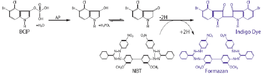
Measurement principle
BCIP, a chromogenic substrate for alkaline phosphatase (AP), produces an indigo color by reaction. In the same reaction, NBT is reduced and turns into an insoluble purple precipitate.
Usage
Procedure for staining endogenous alkaline phosphatase (AP). For other applications of usage, please refer product instruction.
- Fix cells with a fixing solution such as paraformaldehyde
- Wash fixing solution with water.
- Pour BCIP-NBT Solution slowly, and leave at room temperature to react.
(For 35mm dish, please use 1mL of BCIP-NBT Solution) - When an appropriate stained image is obtained, wash with water to stop the reaction.
- Observe it enclosed or after air drying.
Ordering information
| Product Name | Grade | Storage | Catalog Number | PKG Size | Price |
|---|---|---|---|---|---|
| BCIP-NBT Solution (Ready To Use) | SP | Refrigerate | 19880-84 | 100 mL |
Related product
| Product Name | Grade | Storage | Catalog Number | PKG Size | Price |
|---|---|---|---|---|---|
| Blocking One Histo | SP | Refrigerate | 06349-64 | 50 mL | |
| HistoVT One (10x, pH 7.0) | SP | Room temperature | 06380-76 | 100 mL | |
| 06380-05 | 500 mL | ||||
| 4%-Paraformaldehyde Phosphate Buffer Solution |
SP | Refrigerate | 09154-14 | 5 x 10 mL | |
| 09154-56 | 100 mL | ||||
| 09154-85 | 500 mL |
