
AAT Bioquest社 リソソーム染色キット
明るく退色しにくい色素

Lysosomes are cellular organelles which contain acid hydrolase enzymes to break up waste materials and cellular debris. Lysosomes
digest excess or worn-out organelles, food particles, and engulfed viruses or bacteria. The membrane around a lysosome allows the digestive enzymes to work at pH 4.5. The interior of the lysosome is acidic (pH 4.5-4.8) compared to the slightly alkaline
cytosol (pH 7.2). The lysosome maintains this pH differential by pumping protons from the cytosol across the membrane via proton pumps and chloride ion channels.
is significantly enhanced upon entering lysosomes. This key feature significantly reduces the staining background and makes the assay kits useful for a variety of studies, including cell adhesion, chemotaxis, multidrug resistance, cell viability, apoptosis
and cytotoxicity. The Cell Navigator™ staining kits are suitable for proliferating and non-proliferating cells, and can be used for both suspension and adherent cells. The labeling protocols are robust, requiring minimal hands-on time. The kits can be readily adapted for many types of fluorescence platforms such as microplate
assays, flow cytometry and fluorescence microscope.
Key Features of Cell Navigator™ Staining Kits:
• Minimal cytotoxicity, no cell toxicity observed.
• Multiplexing wavelengths, Ex/Em = 450/505 nm, 542/556 nm, 575/597 nm and 596/619 nm.
• Increased signal intensity, 10 times brighter than the product of Company A.
• Extraordinarily high photostability, no fading observed with 2 minutes exposure.
• Excellent cellular retention, more than 6 passages for cell tracking in Hela cells.
• Fixable, cell staining pattern survives fixation.
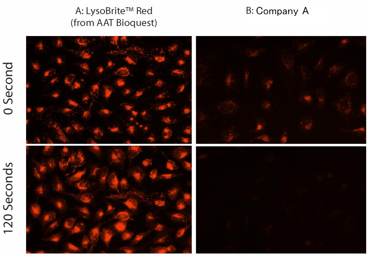
Figure 1. Images of Hela cells stained with A: Cell Navigator™ Lysosome Staining Kit, B: the product from Company A in a Costar blackwall/clear bottom 96-well plate. The signals were compared at 0 and 120 seconds exposure time by using an Olympus fluorescence microscope.
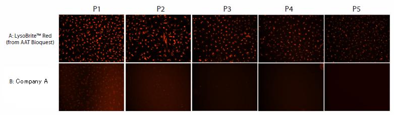
Figure 2. Images of Hela cells stained with A: Cell
Navigator Lysosome Staining Kit (Top, from AAT Bioquest), B: the product from Company A in a Costar black wall/clear bottom 96-well plate. The signals were compared at 5 cell passages (P1, P2, P3, P4 and P5) respectively using an Olympus fluorescence microscope.
| Cat.# | Product Description | Ex(nm) | Em(nm) | Size |
| 22656 | Cell Navigator Lysosome Staining Kit Green Fluorescence | 450 | 505 | 500assay |
| 22657 | Cell Navigator Lysosome Staining Kit Orange Fluorescence | 542 | 556 | 500assay |
| 22658 | Cell Navigator Lysosome Staining Kit Red Fluorescence | 575 | 597 | 500assay |
| 22659 | Cell Navigator Lysosome Staining Kit Deep Red Fluorescence | 596 | 619 | 500assay |
Fount of Information は、新商品、新規取扱メーカーなどの情報をいち早く紹介するコンテンツです。情報発信のスピードを重視しているコンテンツのため、現時点で法規制や取り扱いを確認できていない商品、定価を設定できていない商品があります。ご要望やご照会を受けた商品について、法令整備や在庫の充実を図ります。
