
AAT Bioquest社 アポト-シスとネクローシス検出キット
3種類の色でアポトーシス、ネクローシス、正常細胞を同時検出

Apoptosis is an active, programmed process of autonomous cellular dismantling
that avoids eliciting inflammation. In apoptosis, phosphatidylserine (PS)
is transferred to the outer leaflet of the plasma membrane. As a universal
indicator of the initial/intermediate stages of cell apoptosis, the appearance of
phosphatidylserine on the cell surface can be detected before morphological
changes are observed.
Necrosis is characterized as a passive, accidental cell death resulting from environmental
perturbations with uncontrolled release of inflammatory cellular contents. Loss of plasma membrane integrity represents a straightforward approach to demonstrate late stage apoptosis and necrosis.
AAT Bioquest offers Cell Meter™ Apoptotic and Necrotic Detection Kits as a set of tools for monitoring cell viability. Our Cell Meter™ detection kits are optimized to simultaneously detect cell apoptosis, necrosis and healthy cells with a flow cytometer or fluorescence microscope. The phosphatidylserine (PS) sensor used in Kit 22843 has deep red fluorescence (Ex/Em = 630/660 nm) upon binding to membrane PS. Membrane-impermeable Nuclear Green™ DCS1 (Ex/ Em = 490/525 nm) is used to label the nucleus while CytoCalcein™ Violet 450 (Ex/Em = 405/450 nm) is provided for labeling live cell cytoplasm.
Key Features of Cell Meter™ Detection Kits:
• Multiplexing capability, triple colors for the simultaneous detection of multiple cellular events.
• Robust, a mix and read format.
• Convenient, compatible with common filter sets.
JC-10, a Superior Replacement to JC-1
◊ More water soluble, DMSO stock solution of JC-10 can be readily diluted into a variety of aqueous buffers.
◊ More sensitive, detect smaller mitochondrial membrane potential changes in certain cell lines.
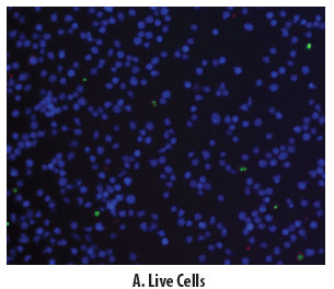
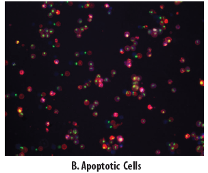
Figure 1. The detection of binding activity of Apopxin™ Deep Red to phosphatidylserine in Jurkat cells. The fluorescence images demonstrated Jurkat cells that are live (blue, stained by CytoCalcein™ Violet 450), apoptotic (red, stained by Apopxin™ Deep Red), and necrotic (green, indicated by Nuclear Green™ DCS1staining). The cells were induced by 1μM staurosporine for
3 hours. The fluorescence images of the cells were taken with Olympus fluorescence microscope through the Violet, Cy5 and FITC channels respectively. Individual images were taken from
each channel for the same cell population. The images were merged as shown above. A: Non-induced control cells; B: Triple staining of staurosporine-induced cells.
|
Cat. # |
Product Description | Ex (nm) | Em (nm) | Size |
| 22840 | Cell Meter™ Apoptotic and Necrotic Detection Kit *Triple Fluorescence Color* | 405 | 450 | 100 Assays |
| 490 | 525 | |||
| 546 | 647 | |||
| 22843 | Cell Meter™ Apoptotic and Necrotic Detection Kit *Triple Fluorescence Color* | 405 | 450 | 100 Assays |
| 490 | 525 | |||
| 630 | 660 | |||
| 22811 | Cell Meter™ Nuclear Apoptosis Assay Kit *Green Fluorescence* | 503 | 526 | 100 Assays |
| 22844 | Cell Meter™ TUNEL Apoptosis Assay Kit | 556 | 579 | 50 Assays |
| 13433 | Z-DEVD-ProRed™ 620 *Red Caspase 3/7 Substrate* | 534 | 619 | 1 mg |
Fount of Information は、新商品、新規取扱メーカーなどの情報をいち早く紹介するコンテンツです。情報発信のスピードを重視しているコンテンツのため、現時点で法規制や取り扱いを確認できていない商品、定価を設定できていない商品があります。ご要望やご照会を受けた商品について、法令整備や在庫の充実を図ります。
