|
Autophagy is a natural and regulated lysosomal degradation pathway essential for cell viability, development and homeostasis. It also serves as an adaptive mechanism to protect organisms against various pathologies.
Decreases and defects in autophagy have been implicated in multiple diseases, for example Huntingtons, Alzheimers, and Parkinsons. In terms of cancer development, autophagy seems to play multiple roles.
To investigate the role autophagy plays in cell homeostasis, imaging tools and assays have been developed to monitor autophagy functionality in response to cellular stress, microbial infection and disease. Autophagy assays, either customized in-house or tested commercially-available kits, employ specific autophagosome markers to analyze the activity of autophagy.
Our Cell Meter™ Autophagy Fluorescence Imaging Kits utilize our proprietary Autophagy Blue™, Green™, & Super Blue™ probes, to selectively target and analyze autophagy activity, which have much higher selectivity than other commercially available autophagy probes.
|
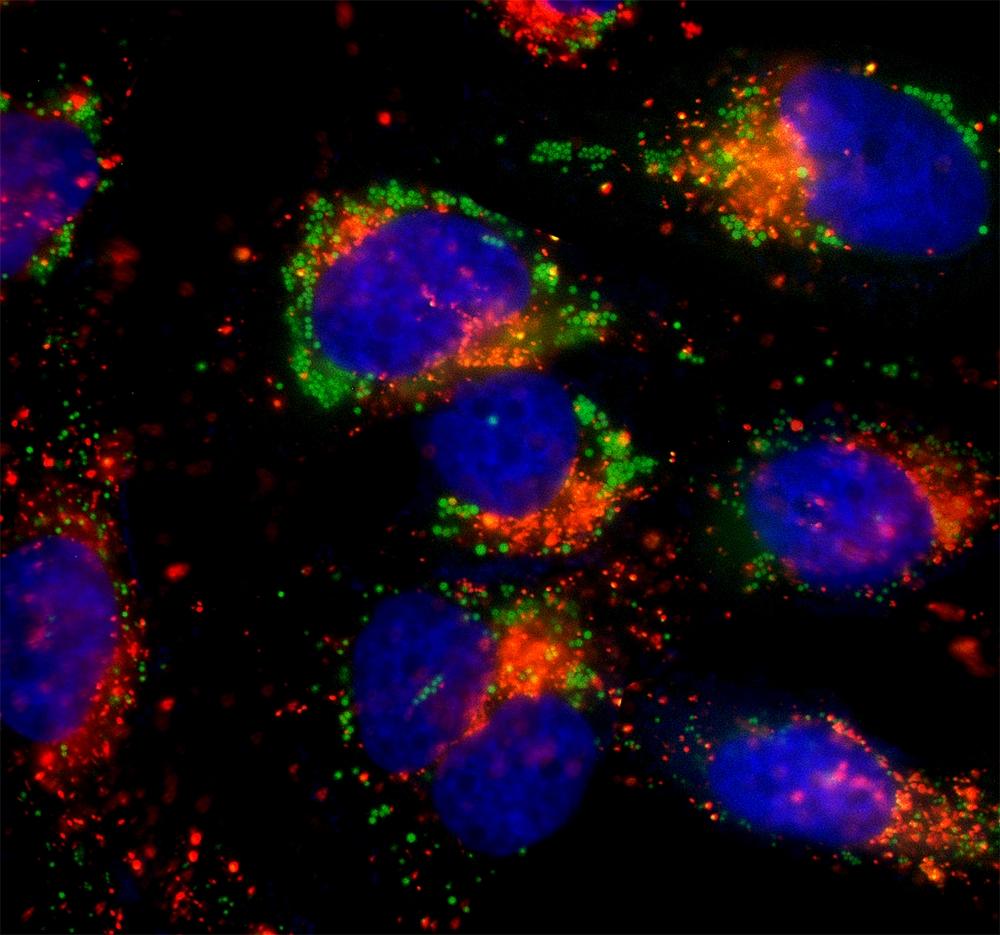
Multicolor Fluorescent Visualization: HeLa cells were incubated in 1X HBSS buffer with 5% serum to induce starvation. Following starvation, cells were treated with Autophagy Green™ (Green) working solution for 20 minutes and then washed 3 times. Nuclei were labeled with Hoechst 33342 (Blue). Lysosomes were labeled with LysoBrite™ Orange (Orange).
Lysosome/Autophagosome
Catalog
|
|
Resources & Information
AAT Bioquest has a wide range of resources and specialized products for researchers, from Application Notes & FAQs to spectral profiles.
Related Digital Catalogs:
• Apoptosis and Necrosis
• Cellular Processes
AssayWise Newsletter Research Articles:
• Multiplex Apoptosis and Necrosis
• NAADP-AM, A New Player in Calcium Signaling Pathways
Customer Favorites
Cell Meter™ Autophagy Kits:
• Autophagy Assay Kit Series
• Autophagy Fluorescence Imaging Kit
• Mitochondrial Autophagy Imaging Kit
|
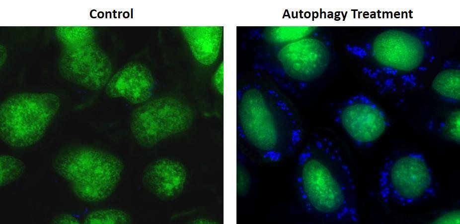
Comparison of Healthy vs. Starved Cells: Autophagy Super Blue™ labeled vesicles were induced by starvation in HeLa cells. HeLa cells were incubated in a regular DMEM medium (Left: Control) or in 1X HBSS buffer with 5% serum (Right: Autophagy Treatment) for 16 hours. Cell nuclei were stained with Nuclear Green™ LCS1 (green).
|

