
AAT Bioquest社 Using Phalloidin's Smaller Size for Improved Actin Labeling Density and Imaging
|
Labeling Actin: Phalloidin vs. Antibody Conjugates |
||
|
Fluorescent-labeled phalloidin is enormously useful in localizing actin filaments in living or fixed cells as well as for visualizing individual actin filaments in vitro. In comparison to antibodies, phalloidin-dye conjugates are smaller, so labeling does not impede cell function. This small size also permits much denser labeling, which allows for more detailed images at higher resolutions.
|
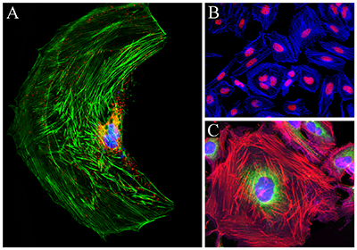 Cell Types & Phalloidin Staining: Cell Types & Phalloidin Staining:A) CPA cell labeled with Phalloidin iFluor® 488, and co-stained with lysosomal dye LysoBrite™ Red and nuclei stain Nuclear Blue™ DCS1. B) HeLa cells labeled with Phalloidin-iFluor® 350 and co-stained with nuclei stain Nuclear Red™ DCS1. C) HeLa cells labeled with Phalloidin-iFluor® 555, and co-stained with mitochondria stain MitoLite™ Green FM, nuclei stain Nuclear Blue™ DCS1, and plasma membrane stain Cellpaint™ Deep Red. |
|
|
Resources & Information
AAT Bioquest has a wide range of resources and specialized products for researchers, from Application Notes to FAQs. |
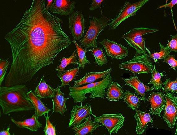 Multicolor Fluorescent Imaging: HeLa cells were fixed in 4% formaldehyde, co-labeled with mitochondria dye MitoLite™ Red FX600 (red), actin filaments with Phalloidin- iFluor® 488 Conjugate (green), and nuclei were stained with Nuclear Blue™ DCS1 (blue). Multicolor Fluorescent Imaging: HeLa cells were fixed in 4% formaldehyde, co-labeled with mitochondria dye MitoLite™ Red FX600 (red), actin filaments with Phalloidin- iFluor® 488 Conjugate (green), and nuclei were stained with Nuclear Blue™ DCS1 (blue). |
|
Fount of Information は、新商品、新規取扱メーカーなどの情報をいち早く紹介するコンテンツです。情報発信のスピードを重視しているコンテンツのため、現時点で法規制や取り扱いを確認できていない商品、定価を設定できていない商品があります。ご要望やご照会を受けた商品について、法令整備や在庫の充実を図ります。
