Cytoskeleton社 F-actin蛍光染色試薬 Acti-stain™ Fluorescent Phalloidins
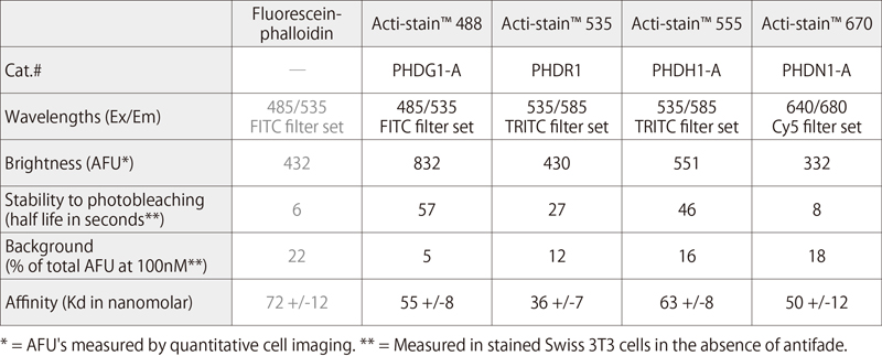
Acti-stain™シリーズは、F-actin(アクチン)の検出に用いられるPhalloidin(ファロイジン)に、高輝度かつ退色耐久性に優れた色素をコンジュゲートした製品です。
詳細情報 >> Cytoskeleton社WEBサイト
メーカー情報
The Acti-stain™ line of fluorescent phalloidins has been developed with an emphasis on creating exceptionally bright and stable probes at an economical price. Side-by-side comparisons with leading competing products demonstrate that you will enjoy savings while not sacrificing the quality of the stain when switching to an Acti-stain™ probe. Additionally, these probes are compatible with popular filter sets such as FITC, TRITC and Cy5. The combination of in-house manufacturing, stringent quality control and convenient packaging provides a great value. Give them a try and see for yourself.
Product Uses Include
- Stain F-actin in fixed cells
- Stabilize actin filaments in vitro
- Visualize actin filaments in vitro
Actin staining is very useful in determining the structure and function of the cytoskeleton in living and fixed cells. The actin cytoskeleton is a very dynamic and labile structure in the living cell, but it can be fixed with paraformaldehyde prior to probing or staining for actin structures.
■ Biochemical characteristics of fluorescent phalloidins
● Acti-stain™ 488
 |
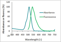 |
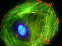
| Swiss 3T3 cell stained with anti-vinculin (red), Dapi (blue nucleus) and F-actin is stained with Acti-stain™ 488 (green F-actin, Cat.# PHDG1). | Absorbance and fluorescence scan of Acti-stain™ 488. Labeled phalloidin was diluted into methanol and its absorbance and excitation spectra were scanned between 350-750 and 500-750 nm, respectively. Absorbance peaks at 500 nm and fluorescence at 550 nm. |
● Acti-stain™ 555
 |
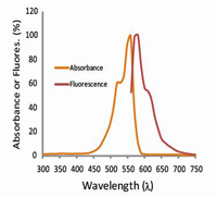 |
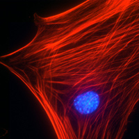
| Swiss 3T3 cell with activated Rho, nucleus is stained with Dapi (blue) and F-actin is stained with Acti-stain™ 555 (red F-actin, Cat.# PHDH1). | Absorbance and fluorescence scan of Acti-stain™ 555. Labeled phalloidin was diluted into methanol and its absorbance and excitation spectra were scanned between 300-750 and 560-750 nm, respectively. Absorbance peaks at 560 nm and fluorescence at 575 nm. |
● Acti-stain™ 670
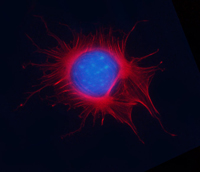 |
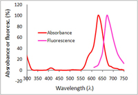 |
| Mitotic Swiss 3T3 cell, F-actin stained with Acti-stain™ 670 (far-red, Cat. # PHDN1), nuclear DNA stained with Dapi (blue). | Absorbance and fluorescence scan of Acti-stain™ 670. Labeled phalloidin was diluted into methanol and its absorbance and excitation spectra were scanned between 300-750 and 600-750 nm, respectively. Absorbance peaks at 625 nm and fluorescence at 675 nm. |
詳細情報 >> Cytoskeleton社WEBサイト
Fount of Information は、新商品、新規取扱メーカーなどの情報をいち早く紹介するコンテンツです。情報発信のスピードを重視しているコンテンツのため、現時点で法規制や取り扱いを確認できていない商品、定価を設定できていない商品があります。ご要望やご照会を受けた商品について、法令整備や在庫の充実を図ります。


