
Molecular Targeting Technologies Inc. 社ホスファチジルセリン検出プローブ
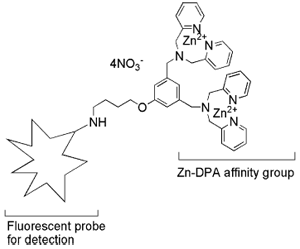
PSVue® はbis(zinc2+dipicolylamine:Zn-DPA)を含む蛍光プローブです。Zn-DPAは陰イオン性リン脂質に高い親和性を示すことが知られており、細胞膜上のフォスファチジルセリン(PS)へ結合します。アポトーシス1、ネクローシス2、グラム陰性/陽性細菌3-5等の検出に使用することができます。。
特長
- UV~近赤外(検出波長)までラインアップ
- in vitroとin vivoに使用可能
- HTSスクリーニングへ適用可能
- アネキシンVのようにPSへ結合
アネキシンVに勝る点
- 結合が早い6,7
- カルシウムイオン非依存的に結合(膜のscramblaseによる影響が無い)8
- 広い範囲の条件(例:10%血清存在下、4~37℃)でアポトーシスを検出8
- 小さい(1つのアネキシンVが結合する範囲に10個以上結合する)9
PSVue®の構造
膜結合の模式図
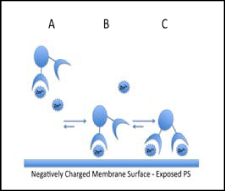
生理条件下では、ほとんどのPSVueはZn2+イオン1分子が結合している(A)。PSVue®は陰イオンのPSへZn2+を介して結合し(B)、もう1分子のZn2+を更に結合させて膜へ結合していると考えられる(C)。
In vitroでの実施例
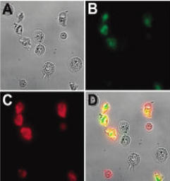
アネキシンVとPSVue®794での多重染色
Jurkat細胞をカンプトテシン10μMで3.5時間処理し、アネキシンV連結色素とPSVue®794両色素で染色した。A:明視野、B:アネキシンV連結色素、C:PSVue®794、D:重ね合わせ
(データ提供:Notre Dame大学 Dr. Bradley Smith)
In vivoでの実施例
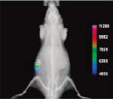
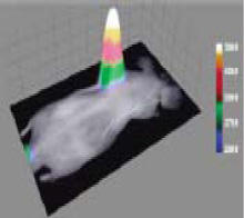
前立腺癌モデルラットでの実施例
PSVue®794(4.0mg/kg)をインジェクションして24時間後に観察したところ、はっきりと癌組織に蓄積していた。
EMT-6移植ヌードマウスでの実施例
PSVue®794をインジェクションして24時間後に観察したところ、はっきりと癌組織に蓄積していた。
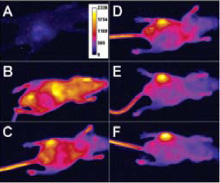
黄色ブドウ球菌感染マウスでの実施例
左後肢の大腿筋へ接種する前(A)、直後(PSVue®794インジェクション)(B)、6時間後(C)、12時間後(D)、18時間後(E)、21時間後(F)
データ提供:Notre Dame大学 Dr. Bradley Smith
参考文献
- Hanshaw RG, Lakshmi C, Lambert TN, Johnson JR and Smith BD. Fluorescent detection of apoptotic cells by using zinc coordination complexes with a selective affinity for membranes surfaces enriched with phosphatidylserine. ChemBioChem, 2005, 6, 2214-2220.
- Smith BA, Akers WJ, Leevy WM, Lampkins AJ, Xiao S, Wolter W, Suckow MA, Achilefu S and Smith BD. Optical imaging of mammary and prostrate tumors in living animals using a synthetic near infrared zinc(II)-dipicolylamine probe for anionic cell surfaces. J. Am. Chem. Soc. 2010, 132 (1), 67-69.
- Leevy, WM; Johnson, JR.; Lakshmi, C.; Morris, J.; Marquez, M.; Smith, BD. Selective recognition of bacterial membranes by zinc(II)-coordination complexes. Chem. Commun. 2006, 595-1597.
- Leevy WM, Gammon, ST, Jiang H, Johnson JR, Maxwell DJ, Marquez M, Piwinica-Worms D and Smith BD. Optical imaging of bacterial infection in living mice using a fluorescent near-infrared molecular probe. J. Am. Chem. Soc., 2006, 128, 16476-16477.
- Leevy WM, Gammon ST, Johnson JR, Johnson JR, Lampkins AJ, Jiang H, Marquez M, Piwinica-Worms D and Smith BD. Noninvasive optical imaging of Straphylococcus aureus bacterial infection in living mice using a bis-dipicolylamine-zinc (II) affinity group conjugated to a near infrared fluorophore. Bioconj. Chem. 2008, 19, 686-692
- DiVittorio KM, Johnson JR, Johansson E, Reynolds AJ, Jolliffe KA and Smith BD. Synthetic peptides with selective affinity for apoptotic cells. Org. Biomol. Chem., 2006, 4, 1966-1976.
- Koulov AV, Stucker KA, Lakshmi C, Robinson JP and Smith BD. Detection of apoptotic cells using a synthetic fluorescent sensor for membrane surfaces that contain phosphatidylserine. Cell Death and Differentiation, 2003, 10, 1357-1359
- Hanshaw RG and Smith BD. New reagents for phosphatidylserine recognition and detection of apoptosis. Bioorg. & Med. Chem. 2005, 13, 5035-5042
- Koulov AV, Hanshaw RG, Stucker KA, Lakshmi C and Smith BD. Biophysical studies of a synthetic mimic of the apoptosis-detecting protein annexin V. Israel. J of Chem., 2005, 45, 373-379.
価格表(構造式の画像をクリックすると拡大してご覧いただけます。)
| 製品名 | メーカー 製品番号 | 構造式 | Ex/Em (nm) | 容量 | オンライン カタログへ |
|---|---|---|---|---|---|
| PSVue® 794 | P-1001 |  |
794/810 | 1kit | |
| キット構成:PSVue® 794(1mMに溶解後約0.68mL)、希釈液、硝酸亜鉛バッファー | |||||
| PSVue®380 | P-1002 |  |
380/440 | 1kit | |
|
キット構成:PSVue®380(2mMに溶解後約0.4mL)、硝酸亜鉛バッファー |
|||||
| PSVue®480 | P-1003 |  |
480/519 | 1kit | |
|
キット構成:PSVue®480(1mMに溶解後約0.5mL)、硝酸亜鉛バッファー |
|||||
| PSVue®Biotin | P-1004 | 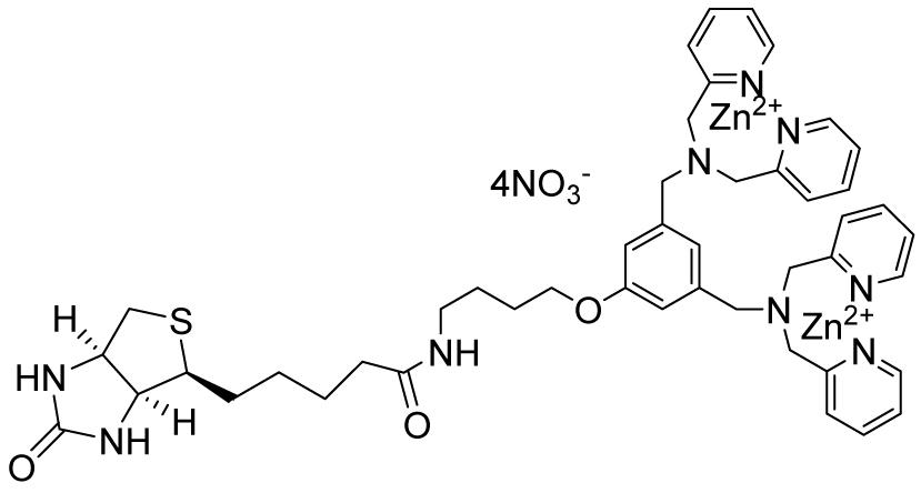 |
- | 1mg | |
|
キット構成:PSVue®Biotin(約1mg)PSVue®Biotinの希釈はTESバッファーを使用します。 |
|||||
| PSVue®550 | P-1005 | 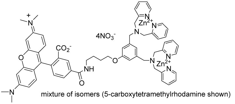 |
553/615 | 1kit | |
|
キット構成:PSVue®550(1mMに溶解後約0.5mL)、硝酸亜鉛バッファー |
|||||
